Birth Weight to Placental Weight Ratio and Left Ventricular Dimensions in Newborns
PROSPECTIVE COHORT STUDY
J. Perinat. Med. 2024; 52(4): 433–444
Published online: March 27, 2024
https://doi.org/10.1515/jpm-2023-0384
Impact of birth weight to placental weight ratio and other perinatal risk factors on left ventricular dimensions in newborns: a prospective cohort analysis
Ashraf Gad, Dhafer Malouche, Manoj Chhabra, Danthanh Hoang, Debbie Suk, Nitin Ron, Beata Dygulska, Madhu B. Gudavalli, Ali M. Nadroo, Pramod Narula, Ibrahim Elmakaty
Abstract
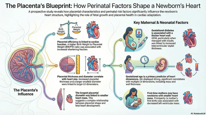
This prospective cohort study investigated the association between birth weight to placental weight (BW/PW) ratio and echocardiographic left ventricle (LV) morphology at birth, while accounting for other relevant perinatal factors. We analyzed 827 neonates at NewYork-Presbyterian Brooklyn Methodist Hospital (2014–2018), categorized by their BW/PW percentile into three groups: small (n=16), normal (n=488), and large (n=323). Key findings reveal that placental thickness and smallest diameter were positively correlated with several LV parameters, while the BW/PW ratio correlated with increased shortening fraction. This study highlights the importance of fetal growth and placental health in the physiological adaptation of the fetal heart.
Key Findings
| Placental Factor | LV Parameter | Effect | p-value |
|---|---|---|---|
| Placental thickness | IVSd | Positive correlation | p=0.002 |
| Placental thickness | IVSs | Positive correlation | p=0.001 |
| Placental thickness | LVPWd | Positive correlation | p=0.003 |
| Placental thickness | LVPWs | Positive correlation | p<0.001 |
| Placental thickness | LV mass | Positive correlation | p=0.017 |
| Smallest diameter | Multiple LV parameters | Positive correlation | p<0.001 |
| BW/PW ratio | Shortening fraction | Estimate=0.29 (95% CI 0.03–0.55) | p=0.027 |
| Longest diameter | LVIDd | Decrease | p=0.039 |
| Longest diameter | LV mass | Decrease | p=0.024 |
Study Highlights
- Population: 827 neonates categorized into three BW/PW ratio groups
- Design: Single-center prospective cohort study with echocardiography within 48–72 hours after delivery
- Statistical Methods: Multiple imputations with PCA, genetic algorithm for model selection, backward stepwise regression
- Model Fit: LV mass model showed highest R² (0.406), indicating strong relationship with predictor variables
Clinical Implications
- Placental dimensions independently affect LV morphology in newborns
- BW/PW ratio serves as a valuable measure of placental efficiency
- Gestational age strongly correlates with multiple LV dimensions
- Maternal factors (GDM, insulin use, primiparity) influence neonatal cardiac development
- Findings may inform early cardiovascular risk assessment strategies
Regression Model Results
| LV Parameter | R² | Key Predictors |
|---|---|---|
| LV mass | 0.406 | GA, placental dimensions, sex, size category |
| IVSd | 0.341 | GA, placental diameter, thickness, NICU admission |
| IVSs | 0.302 | GA, ponderal index, placental factors, GDM |
| LVIDd | 0.266 | GA, chest circumference, maternal BMI, sex |
Dashboard Features
- Interactive visualizations of regression outcomes and correlation matrices
- Pathway diagrams showing placental → cardiac relationships with directional arrows
- Subgroup analysis toggle between small, normal, and large BW/PW groups
- Hover tooltips displaying regression coefficients, p-values, and confidence intervals
- Significance heatmap for predictor-outcome associations
Citation
Gad A, Malouche D, Chhabra M, Hoang D, Suk D, Ron N, Dygulska B, Gudavalli MB, Nadroo AM, Narula P and Elmakaty I (2024) Impact of birth weight to placental weight ratio and other perinatal risk factors on left ventricular dimensions in newborns: a prospective cohort analysis. J. Perinat. Med. 52(4): 433–444. doi: 10.1515/jpm-2023-0384